Prostate biopsy
Depending on the results of the physical (DRE) examination or PSA test, your doctor may refer you to hospital to have a biopsy taken.1,4
A biopsy is when samples of tissue from the prostate gland are taken for further analysis.1 The results should confirm whether prostate cancer is present or not.
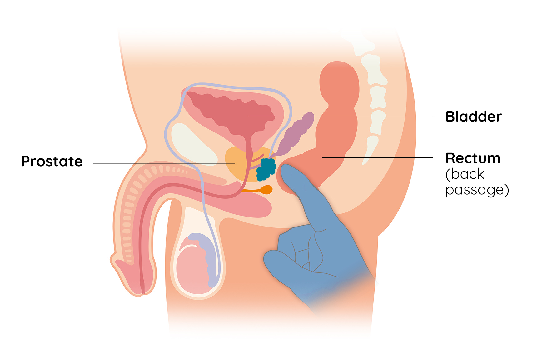
Transrectal ultrasound scan guided (TRUS) biopsy
A slim ultrasound probe is inserted into the back passage (rectum). This creates images that enable the operator to see where to place a needle and take 10-12 samples of prostate tissue.1,4 Since this can be uncomfortable or painful, a local anaesthetic may be given.3 Antibiotics may also be needed to reduce the risk of infection.1,4
Transperineal biopsy
A transperineal biopsy is usually performed under general anaesthesia.4 This involves a needle going into the skin behind the scrotum.4 Again, a few tissue samples will be obtained that can then be examined under the microscope to look for cancer cells.4
What happens to the biopsy tissue?
The tissue obtained from the prostate biopsy will be looked at under the microscope. Cells within the tissue will be graded according to how similar they look when compared with healthy prostate cells.1
The grading system for prostate cancer is called the Gleason grading system.1 Under this system cells are given a Gleason grade between 1 and 5, where 1 indicates the cells look normal and 5 indicates that they are abnormal.
An overall Gleason score is calculated based on all the tissue samples obtained during the biopsy. The higher the Gleason score, the more likely the prostate cancer will grow and spread.1